


Type Specific F-Multiplex-HPV Typing Kit

Main Features
- Samples to result in 4 hours
- Ready-to-run mix, just add sample
- Suitable for different samples such as fresh or paraffin embedded tissues (biopsies), liquid-based samples e.g thin prep, ano-genital and oral swab samples
- Differentiates between persistent and new infections
- IVD/CE certified under 98/79/EC
- Manufactured to ISO 9001:2000 ISO 13485:2003
- Optimised to work on Applied Biosystems sequencers
- Free result-evaluation software tools for simple analysis
Copyright F-HPV 2007
f
HPV typing
MULTIPLEX-FLUORESCENT PCR

Click here to Download . the F-HPV Brochure
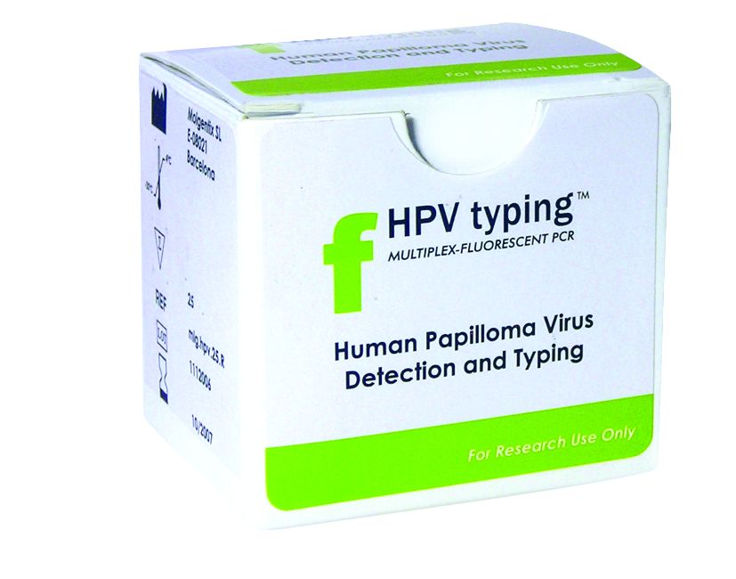


F-HPV typing Multiplex Fluorescent-PCR Kit For Human Papilloma Virus (HPV) Genotyping
Cervical cancer is the second most common cancer causing death in women with an annual incidence of around half a million and a mortality of almost 50%.
The most important risk factor for the development of cervical cancer is genital infection with human papillomavirus (HPV). More than 100 types are known, of which at least 30 usually infect the anogenital tract. HPVs are classified into low and high-risk categories based on their association with malignant lesions. The low-risk types include HPV-6, -11, -42 and -44, and mostly cause the development of genital condylomata (warts).
The high-risk types HPV-16, -18, -31, -33, -35, -39, -45, -51, -52, -56, -58, -59 and -68, induce cervical squamous intraepithelial lesions (SIL), which in turn are classified in low (LSIL) and high grade (HSIL) in severity, and which may progress to cervical cancer. In fact these 13 high-risk HPV types have been shown to be present in up to 99.7% of cervical cancer samples, the most frequently detected being HPV-16, followed by HPV-18, -31, -51, and -45.
Several studies have shown that a persistent HPV infection with the same high-risk type is a risk factor for the development and progression of lesions. If no high-risk specific HPV is present in a cervical sample, the patient has a very low risk to develop cancer for some years. However, if a patient treated of CIN has another positive HPV test within six months after treatment, it will be more predictive of a risk of recurrence for the patient.
On the other hand, women with more than one high-risk type HPV present at the cervix are more likely to have a persistent infection, and are at a strongly increased risk of developing cervical cancer. Therefore, distinguishing between low or high-risk HPV genotypes and especially knowing the specific type is important in the prognostic and follow-up because this permits pinpointing patients with an increased risk for the disease.
F-HPV typing uses 15 primers amplifying within E6 and E7 regions of the HPV genome, the most likely to be retained after viral integration. Extracted DNA is amplified using a multiplex PCR with a set of 16 fluorescently labelled primers recognising HPV types 6, 11, 16, 18, 31, 33, 35, 39, 45, 51, 52, 56, 58, 59, 68 and a human STR used as internal control. This sequence is added to check DNA integrity and PCR inhibitors; it is also helpful in detecting DNA mixtures due to sample mishandling. Different labelling of primers allows to generate amplicons of similar size in the same PCR reaction.